CLADOENDESIS OF EPHEMEROPTERA


|
CLADOENDESIS OF EPHEMEROPTERA |
||||
|
|
zzz | ||
|
|
|
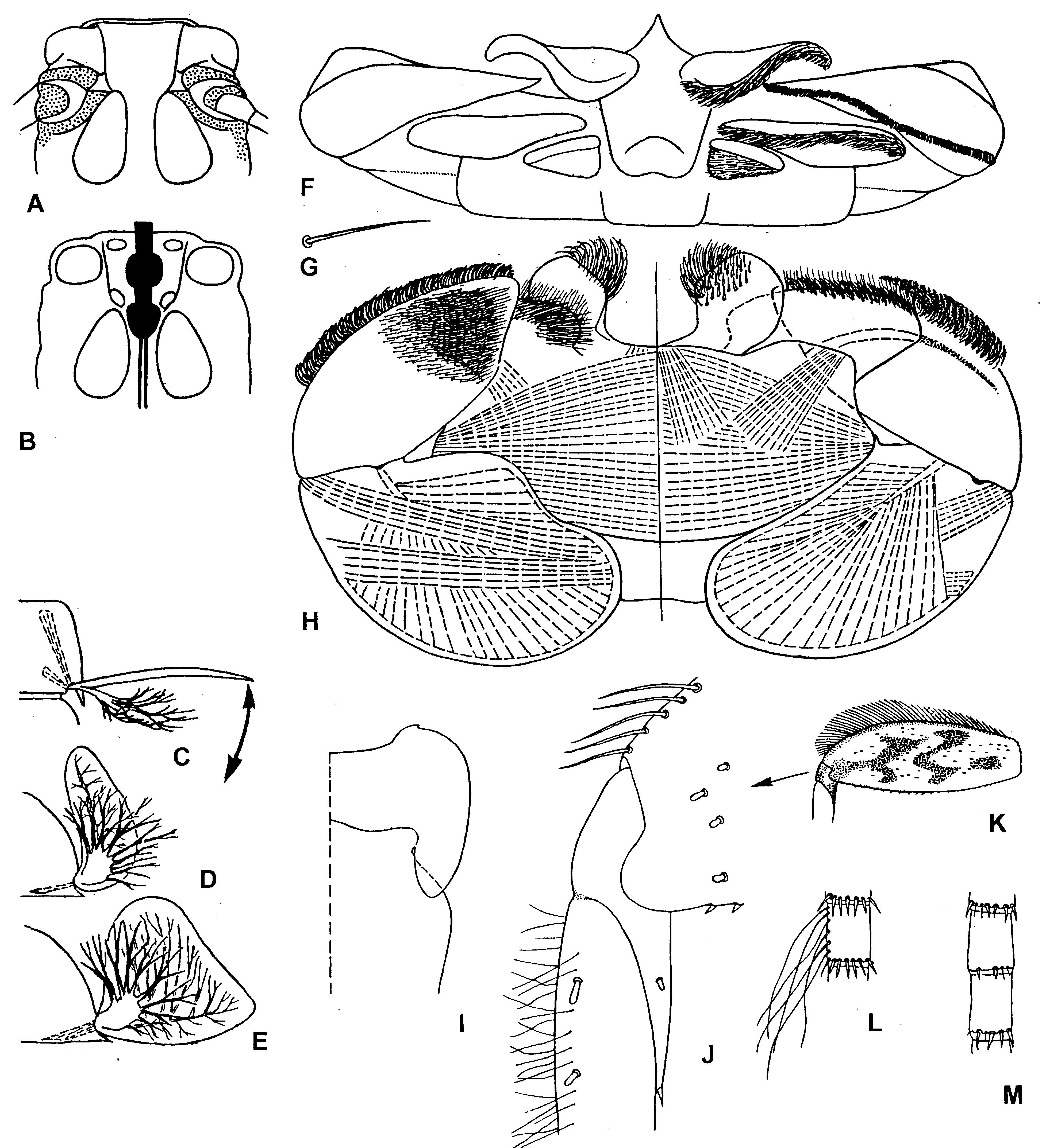
Figure 57. Ecdyonurus/fg1.
A–B – Ecdyonurus/fg1-Afronurus/g1 levis [Epeorus]: A – imaginal mesothorax, ventral view; B – meso- and metathoracic nerve ganglia (shown by black) and ventral areas of mesothoracic muscles attachment (compare with Fig.65:C–D). C–E – scheme of natural position of tergalii in Ecdyonurus/fg1 (muscles shown by interrupted lines) (compare with Fig.61:E–F and 62:H–I): C – dorsal view; D – tergalius I, posterior view; E – tergalius II, posterior view. F–I – Ecdyonurus/fg* venosa [Ephemera]: F – superlinguae, hypopharynx and labium, apical view; G – seta of distal row on dorsal surface of distal segment of labial palp; H – labium, dorsal view (in left half) and ventral view (in right half) (muscles shown by interrupted lines) (compare with Fig.62:E–G); I – left half of larva pronotum and anterior part of mesonotum. J–K – Ecdyogymnurus/g1 scalaris [Ecdyonurus], larval middle right leg, dorsal view: J – apex of femur, patella and base of tibia; K – femur. L – Afghanurus/g* vicinus [Afghanurus], portion of larval cercus. M – Ecdyogymnurus/g1 inversus [Ecdyonurus], the same. (A–H – from Kluge 1988a and 1993a; I–K – from Kluge 1997d; L–M – from Kluge 1980).